0
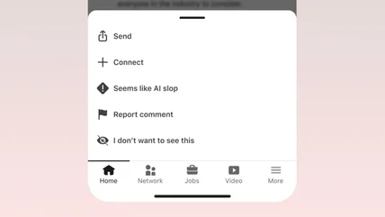
Reddit's CEO sure seems frustrated with Google's AI overviews
Amid rumors of Google breakup, Huffman says "people don't want a summary of reddit, they want reddit."
Read More
Amid rumors of Google breakup, Huffman says "people don't want a summary of reddit, they want reddit."
Read MoreGive me a mixtape, and hold the tape.
Read MoreThe new Friend pendant features a speaker, updated models and the same off-putting ideas about how humans should use AI.
Read MoreThe company is promising the first in-depth look at the $2,195 glasses.
Read MoreUnsurprisingly, it’s likely to focus on AI.
Read MoreWhen it’s less about the product and more about the FCC.
Read MoreGoogle's next foldable doesn't look all that different, save for the camera bar.
Read MoreDespite the slump, Apple and Samsung have both seen their market share go up.
Read MoreThe company is also removing some of its own AI-writing features.
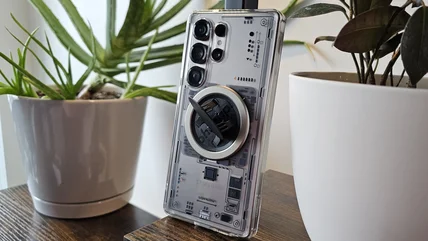
Read MorePhone cases vary dramatically when it comes to pricing, but it's not always as simple as "you get what you pay for."
Read MoreBloomberg says ChatGPT and Roblox will soon be added to the EU’s ‘very large online platform’ list, which subjects them to stricter rules.
Read MoreGoogle said a July update resolves the problem on recent devices, with Pixel 9a and 8a getting a fix in September.
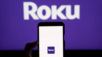
Read MoreYour Roku can probably display what's on your phone screen, if you have Android.
Read MoreThe company is making a big push into fitness, having already partnered with Peloton earlier this year.
Read MoreWith its latest Smart Play sets, Lego managed put its own spin on one of the world's most popular franchises.
Read MoreIf you're looking for a high-performance PC with that clean 'zero-cable' look, Maingear's new pre-built lineup that may suit you.
Read MoreNASA sent a spacecraft called LINK to rescue a falling telescope, but it’s now unclear if it can accomplish its mission.
Read MoreDeepMind's award-winning AlphaFold team is no more.
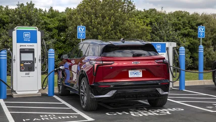
Read MoreElectrified vehicle sales hit new highs around the world as 50 countries saw quarterly records in Q2 of 2026.
Read MoreSamsung's mobile and chip divisions are going in opposite directions, profit-wise.
Read MoreThe program, ChatGPT for Academic Researchers, will start with 10,000 participants this summer.
Read MoreWaymo paused highway operations in May after several robotaxis drove into sections that were closed for construction.
Read MoreMoving your data to Claude is easier than you might think. This step-by-step guide will get you switched over right away.
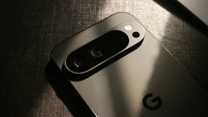
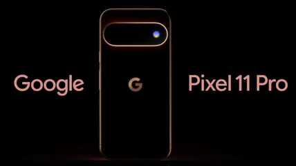
Read MoreWe played spot the difference and noticed this intriguing difference in Google's teaser for the Pixel 11 Pro.
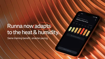
Read MoreIf it's extra hot or humid where you are, the Runna app will adjust your workouts accordingly.
Read MoreX and the World Federation of Advertisers say they're resetting their relationship.
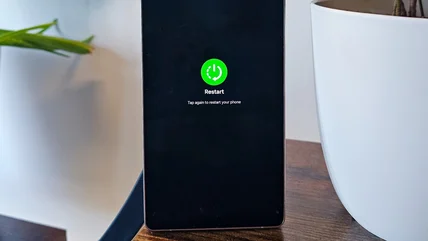
Read MoreIt's a widely held belief that restarting your phone improves performance. Here's the truth.
Read More